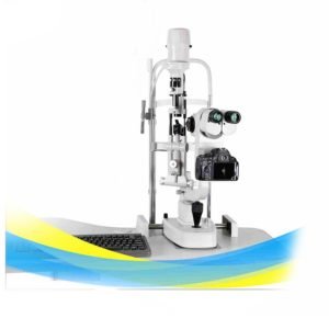
Slit lamp Bio-microscopy NEU3ER-E
£9,950.00
– Advanced Slit Lamp Bio-microscopy NEU3ER-E for precise eye examinations
– Galileo Parallel microscope design for enhanced visibility
– High Definition image capturing with Nikon D 5200 (24.1 Mega pixels)
– Adjustable eyepiece magnification at 12.5x
– Total magnification options: 6x, 10x, 16x, 25x, and 40x
– Diopter adjustment range: -5D to +5D for personalized focus
– Adjustable slit width from 0 to 14mm for versatile applications
– Continuous slit height range of 1 to 14mm for varying conditions
– Slit angle adjustable from 0 to 180 degrees for optimal positioning
– Multiple light spot diameter options: 0.2mm, 1mm, 2mm, 5mm, 10mm, 14mm
– Equipped with various filters: Heat Absorption, Grey, Redfree, and Cobalt Blue
Slit Lamp Bio-microscopy NEU3ER-E
Advanced slit lamp bio-microscopy system delivering precise anterior segment examination with Galileo parallel optics and high-definition image capture. Comprehensive magnification range and adjustable illumination parameters enable detailed clinical assessment across diverse ophthalmic conditions.
Clinical overview
The NEU3ER-E slit lamp bio-microscopy system represents a comprehensive diagnostic platform for detailed anterior segment evaluation. Employing Galileo parallel microscope optics, the system delivers enhanced stereoscopic visualization essential for identifying subtle pathology across the cornea, lens, anterior chamber, and conjunctiva. The integrated high-definition imaging capability via Nikon D5200 (24.1 megapixels) enables clinical documentation and longitudinal comparison, supporting evidence-based patient management and educational applications.
Clinicians benefit from extensive magnification flexibility (6x to 40x), adjustable diopter compensation (−5D to +5D), and versatile illumination parameters including variable slit width (0–14mm), continuous slit height (1–14mm), and rotatable slit angle (0–180°). Multiple light spot diameters (0.2mm to 14mm) and specialized filters (heat absorption, grey, red-free, cobalt blue) facilitate targeted examination of specific anterior structures and enhance contrast for vascular and pigmented lesions.
Key features & surgeon benefits
Galileo Parallel Microscopy
- Enhanced stereoscopic depth perception for precise pathology localization
- Superior image contrast and clarity across all magnification levels
- Reduced optical aberrations for consistent diagnostic accuracy
Versatile Magnification Options
- Five magnification settings (6x, 10x, 16x, 25x, 40x) for comprehensive examination
- Continuous diopter adjustment (−5D to +5D) for personalized focus compensation
- Accommodates diverse refractive errors and clinical requirements
Adjustable Slit Parameters
- Variable slit width (0–14mm) and continuous height (1–14mm) for targeted illumination
- Rotatable slit angle (0–180°) enables optimal positioning for all anterior structures
- Multiple light spot diameters (0.2mm to 14mm) support focused and diffuse examination modes
High-Definition Image Capture
- Integrated Nikon D5200 DSLR (24.1 megapixels) for clinical photography
- Supports longitudinal documentation and patient education
- Facilitates peer review and quality assurance protocols
Specialized Optical Filters
- Heat absorption filter protects anterior segment tissues during prolonged examination
- Grey filter reduces glare and enhances contrast for general evaluation
- Red-free and cobalt blue filters optimize visualization of vascular and pigmented pathology
Intuitive System Design
- Ergonomic positioning and control interfaces minimize clinician fatigue
- Rapid filter and magnification changes support efficient patient throughput
- Seamless integration with standard ophthalmic examination protocols
Technical specifications
| Microscope Type | Galileo Parallel |
|---|---|
| Image Capture | Nikon D5200 DSLR, 24.1 megapixels, high-definition |
| Eyepiece Magnification | 12.5x |
| Total Magnification Options | 6x, 10x, 16x, 25x, 40x |
| Diopter Adjustment Range | −5D to +5D |
| Slit Width | 0–14mm (continuous) |
| Slit Height | 1–14mm (continuous) |
| Slit Angle | 0–180° (adjustable) |
| Light Spot Diameter Options | 0.2mm, 1mm, 2mm, 5mm, 10mm, 14mm |
| Optical Filters | Heat absorption, grey, red-free, cobalt blue |
| Indications | Anterior segment examination, corneal evaluation, lens assessment, anterior chamber analysis |
| Packaging | As applicable |
| Warranty | 2 Year Manufacturer Warranty |
Standard instrument composition
| Item | Description | Qty |
|---|---|---|
| Slit Lamp Microscope Unit | Complete Galileo parallel optical system with integrated illumination | 1 |
| Eyepiece Assembly | 12.5x magnification with diopter adjustment mechanism | 1 |
| Slit Illumination Module | Variable width, height, and angle adjustment system | 1 |
| Light Source | Integrated illumination with spot diameter options | 1 |
| Filter Set | Heat absorption, grey, red-free, and cobalt blue filters | 1 set |
| DSLR Camera Interface | Nikon D5200 mounting and integration system | 1 |
| Chin Rest and Forehead Support | Patient positioning and stabilization assembly | 1 |
| Microscope Stand | Adjustable height and positioning base | 1 |
Get in touch for pricing & availability
Contact our ophthalmic equipment specialists to discuss system configuration, pricing, and delivery timelines. We provide comprehensive support for installation, training, and ongoing clinical optimization.
Email: info@neuvar.co.uk